Cryo-electron Tomography for Cell Biology
In this webinar, you will learn:
– The complete workflow for in situ cryo-electron tomography
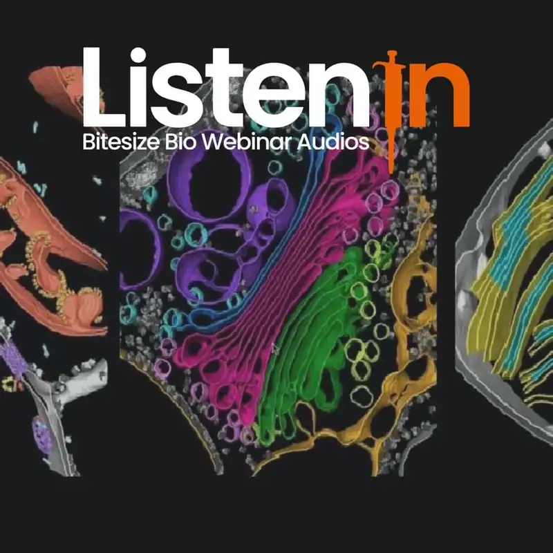
– How subtomogram averaging within the cell yields native-state structures of macromolecular complexes (e.g., the asymmetric and dilated nuclear pore of algae)
– How mapping these structures back into the native cellular environment reveals new molecular interactions that are only accessible by this technique (e.g., the binding of cargo to COPI-coated Golgi membranes and the tethering of proteasomes to the nuclear pore).
Cryo-electron tomography can visualize macromolecular structures in situ, inside the cell. Vitreous frozen cells are first thinned with a focused ion beam and then imaged in three dimensions using a transmission electron microscope. This transformative method has the power to revolutionize our understanding of cell biology, revealing native cellular architecture with molecular clarity.
for more information visit: https://bitesizebio.com/webinar/cryo-electron-tomography-for-cell-biology/
Creators and Guests

Guest
Dr. Ben Engel
Max-Planck-Institute of Biochemistry, Research Department Molecular Structural Biology.


